How to Prepare for a Sheep Brain Dissection: A Guide for Students
Preparing for a sheep brain dissection can be a daunting task for students, but with the right knowledge and materials, the process can become easier. This guide will provide an overview of the steps to take in order to ensure the dissection goes smoothly and safely.
First, it is important to gather all the necessary materials. This includes protective eyewear, a lab coat, gloves, a scalpel, forceps, scissors, and a dissecting tray. Additionally, students should have a reference guide or diagrams of a sheep brain that can be used for comparison during the dissection.
Once the materials have been gathered, students should prepare the area. This includes setting up a lab bench with a dissecting tray, ensuring that the tray is filled with preservative solution, and arranging the tools in an orderly fashion.
[toc]
Next, students should take the time to familiarize themselves with the sheep brain before beginning the dissection. This can be done by looking at the diagrams or reference guide, as well as examining the sheep brain itself. This will help the student to become familiar with the structure and anatomy of the brain, which will be very helpful during the dissection.
Once the student is familiar with the brain, it is time to begin the dissection. To do this safely and effectively, students should wear protective eyewear, as well as a lab coat and gloves. When handling the sheep brain, it is important to use the scalpel and forceps with care and precision. Students should slowly make incisions, taking care to avoid damaging the brain.
Finally, students should clean up the area and dispose of the materials properly. They should also take the time to review their work and compare it to the diagrams to ensure they have accurately dissected the sheep brain.
By following these steps, students can ensure that their sheep brain dissection is successful and safe. With the right preparations and materials, a dissection can be an engaging and informative experience.
Understanding the Anatomy of the Sheep Brain Through Dissection
The sheep brain is a fascinating and intricate organ, composed of many varied parts each with its own purpose. Gaining an understanding of these parts can be achieved through the process of dissection.
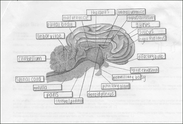
The sheep brain is divided into three main parts: the cerebrum, the cerebellum, and the brainstem. The cerebrum is the largest part of the brain and is responsible for higher functions such as thought, memory, language, and complex motor skills. It is further divided into two hemispheres – the right and the left – that are connected by the corpus callosum.
The cerebellum is located underneath the cerebrum and is responsible for the coordination of movement as well as balance. It is a small, tightly folded structure with two hemispheres.
The brainstem is located below the cerebellum and is responsible for the control of autonomic functions such as breathing and digestion. Its most visible parts are the medulla, the pons, and the midbrain.
When dissecting the sheep brain, it is important to start with a thorough understanding of the anatomy of the brain. An understanding of the major structures and the functions that each of them serves is essential in order to properly identify the different parts.
The first step in the dissection process is to remove the meninges, the protective sheaths that cover the brain. The dura mater is the outermost layer and it is followed by the arachnoid mater and the pia mater. Care should be taken to ensure that these layers are not damaged during the procedure.
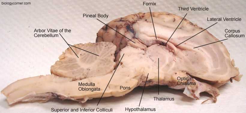
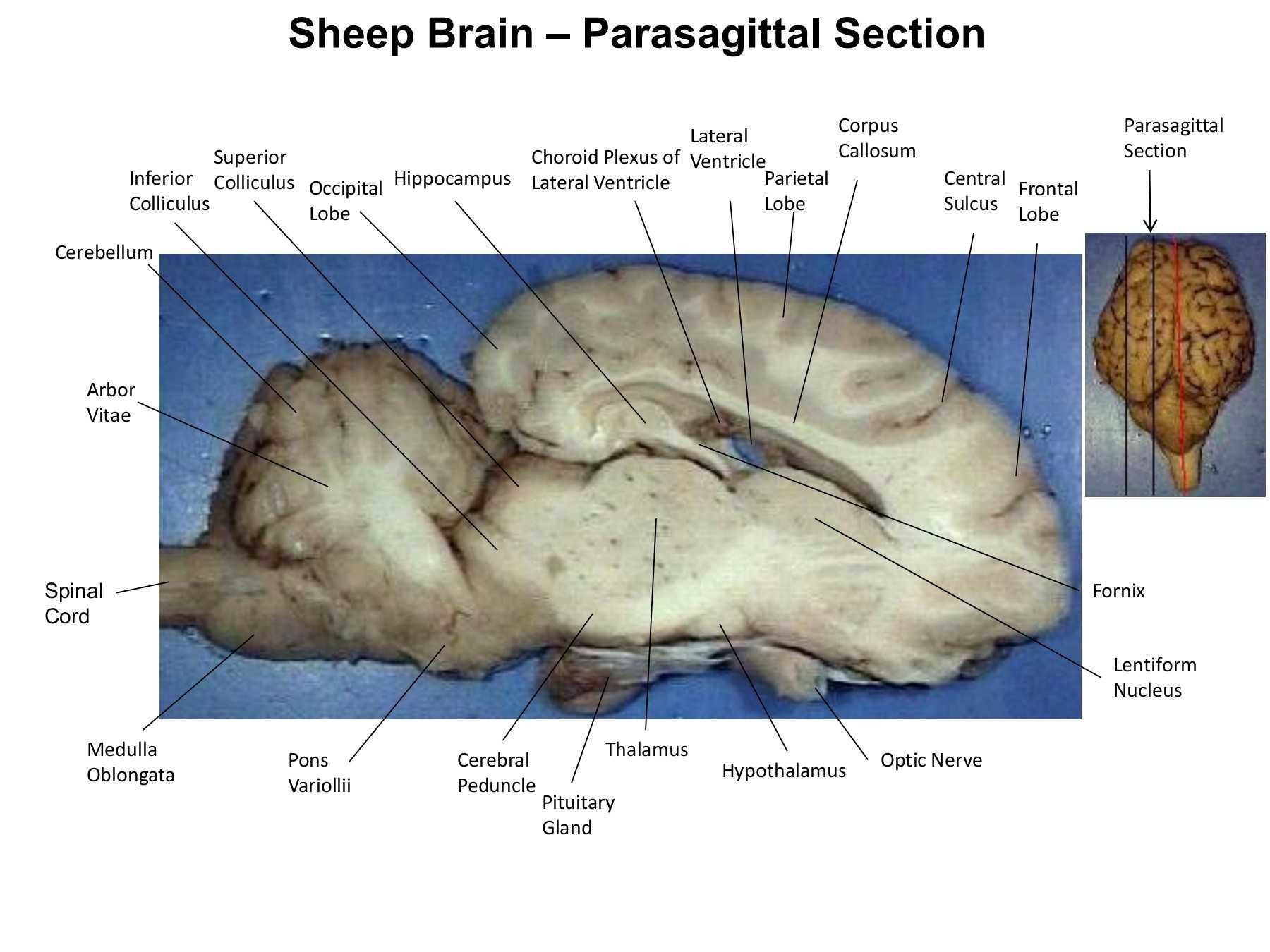
Once the meninges have been removed, the major structures of the brain can be identified and examined. The cerebrum should appear as a large, curved structure with clearly defined hemispheres. The cerebellum is located underneath the cerebrum and is much smaller in size. The brainstem can be identified by its three main components: the medulla, the pons, and the midbrain.
After the major structures of the brain have been identified, the individual parts can be further explored. The major sulci and gyri of the cerebrum should be visible once the dura mater has been removed. The lobes of the cerebrum and the various parts of the cerebellum can also be identified.
Through dissection, one can gain a thorough understanding of the anatomy of the sheep brain. By studying the different parts and their functions, one can gain a better insight into the complex and intricate workings of this vital organ.
Exploring Neuroanatomy Through Sheep Brain Dissection Worksheets
The sheep brain dissection is a powerful tool for exploring neuroanatomy. During the process, students gain an understanding of the structures and functions of the brain and gain an appreciation of the complexity of the nervous system.
The dissection begins by identifying the major structures of the brain. Students remove the meninges, or covering of the brain, and use diagrams to identify the cerebral hemispheres, diencephalon, brainstem and cerebellum.
After the major structures are identified, students use dissection worksheets to explore the details of the brain in greater depth. Students use a scalpel to carefully dissect each structure, removing the connective tissue to reveal the features of the brain. Through this process, students become familiar with the anatomy of the brain and gain an understanding of the various functions of each structure.
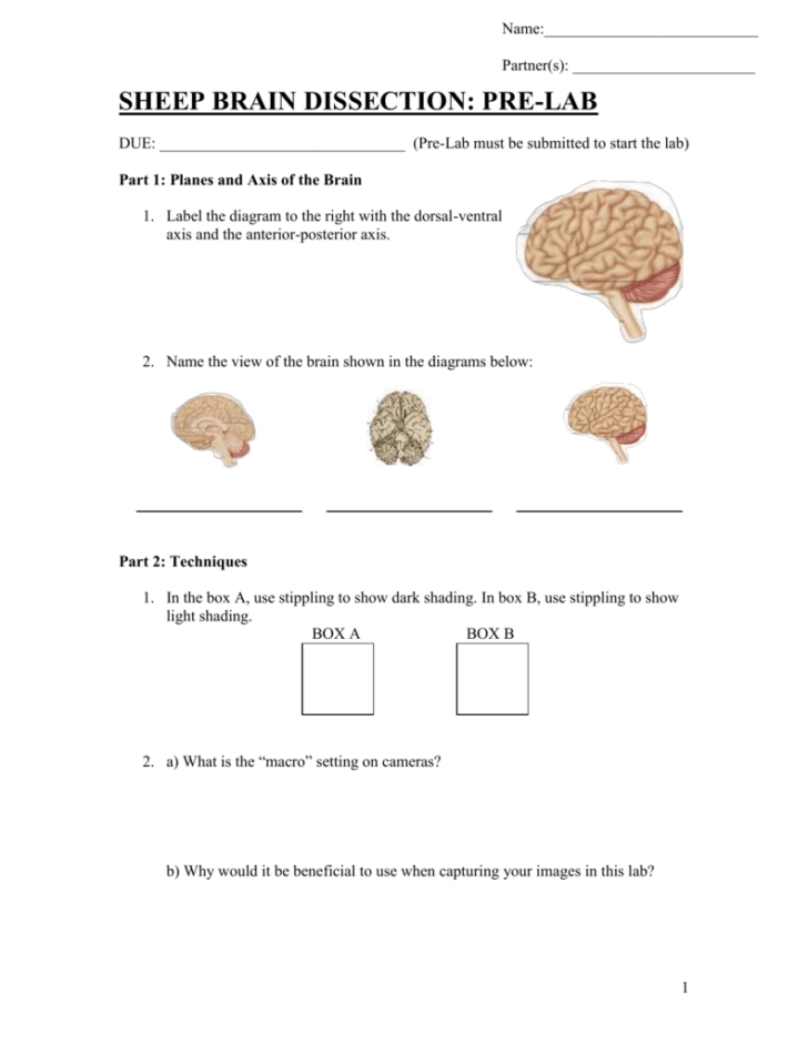
The worksheets are designed to guide the student through the entire process, from the identification of the major structures to the details of the individual components. The worksheets provide an organized approach to the dissection, allowing students to focus on the details without becoming overwhelmed. Each worksheet contains a diagram, anatomical terminology and directions for the student to follow.
By using these worksheets, students gain an appreciation of the complexity of the nervous system. They also gain an understanding of the anatomy of the brain and its various structures and functions. Through the dissection process, students gain a deeper understanding of the brain and its role in the body.
Learning the Parts of the Sheep Brain: An Interactive Worksheet Guide
Welcome to the interactive worksheet guide to learning the parts of the sheep brain. This guide will provide an overview of the various parts of the sheep brain, as well as some basic information about each part.
The sheep brain is divided into four main regions: the cerebrum, the diencephalon, the brain stem and the cerebellum.
The cerebrum is the largest part of the sheep brain and is responsible for the majority of the brain’s functions. It is divided into two hemispheres, the left and the right. Each hemisphere is further divided into four lobes, which are the frontal lobe, the parietal lobe, the temporal lobe and the occipital lobe.
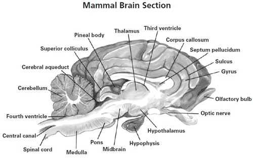
The diencephalon is the region of the brain responsible for things such as regulating body temperature, controlling hunger, and regulating emotions. It is composed of several structures, including the thalamus, the hypothalamus, and the pineal gland.
The brain stem is the part of the brain that connects the cerebrum to the spinal cord. It is responsible for several basic functions, such as controlling heart rate, breathing, and reflexes. It is composed of several structures, including the medulla oblongata, the pons, and the midbrain.
The cerebellum is located at the back of the brain and is responsible for coordinating movement, balancing, and maintaining posture. It is composed of several structures, including the vermis and the hemispheres.
Now that you have a basic understanding of the various parts of the sheep brain, it is time to put your knowledge to the test. Please take the time to complete the activity below to further your understanding of each part of the sheep brain.
Activity:
1. Name the four main regions of the sheep brain.
Answer: The four main regions of the sheep brain are the cerebrum, the diencephalon, the brain stem, and the cerebellum.
2. What is the primary function of the cerebrum?
Answer: The primary function of the cerebrum is to process and integrate sensory information, direct motor functions, and control higher mental functions such as language, memory, and problem-solving.
3. What structures make up the diencephalon?
Answer: The structures that make up the diencephalon are the thalamus, the hypothalamus, and the pineal gland.
4. Name two functions of the brain stem.
Answer: Two functions of the brain stem are controlling heart rate and breathing, and controlling reflexes.
5. What is the primary function of the cerebellum?
Answer: The primary function of the cerebellum is to coordinate movement, balance, and maintain posture.
Conclusion
In conclusion, the Sheep Brain Dissection Worksheet provides a comprehensive and detailed overview of the anatomy and physiology of the sheep brain. It is an excellent resource for students and teachers alike to use in order to gain a better understanding of the structure and function of the sheep brain. The worksheet also provides an excellent opportunity to gain hands-on experience in dissection, allowing students to gain a deeper understanding of the structure of the sheep brain.
[addtoany]
5 photos of the "Sheep Brain Dissection Worksheet"
Related posts of "Sheep Brain Dissection Worksheet"
Incredible Human Machine Worksheet
Exploring the Educational Benefits of the Incredible Human Machine WorksheetThe Incredible Human Machine is an innovative teaching tool that provides an interactive and educational experience for students of all ages. This unique program uses an array of 3D animations and interactive activities to explore the human body, helping students to gain a better understanding of...

Logarithmic Equations Worksheet With Answers
Exploring Logarithmic Equations: A Step-by-Step Guide to Solving Logarithmic Equations Using a Worksheet with AnswersLogarithmic equations are an important part of advanced mathematics and can be used to solve a variety of problems. They are most commonly used to solve exponential equations and can be used to calculate the rate of change of certain values...
Nucleic Acids Worksheet Answers
Exploring the Structure and Function of Nucleic Acids: A Comprehensive Guide to the Nucleic Acids Worksheet Answers1. What are nucleic acids? Nucleic acids are large biologically important molecules that are found in all living organisms. They are composed of nucleotides, which are made up of a sugar, a phosphate group, and a nitrogenous base. Nucleic...
Kinetic Molecular Theory Worksheet
Exploring the Basics of Kinetic Molecular Theory: A Guide to Understanding the Worksheet.Kinetic molecular theory is a fundamental concept in the field of physical chemistry. Put simply, it is an explanation of the behavior of all matter at the atomic and molecular levels. The theory helps us to understand the unique properties of different substances,...