Exploring the Basics of Karyotype Analysis: A Comprehensive Guide to Interpreting Karyotype Worksheet Answers Key
Introduction
Karyotype analysis is a crucial tool for medical professionals to assess the health of a patient. By analyzing the number, shape, and size of chromosomes, medical professionals can gain insight into possible genetic disorders and the health of the patient. In this comprehensive guide, we will explore the basics of karyotype analysis to help interpret karyotype worksheets.

Overview of Karyotype Analysis
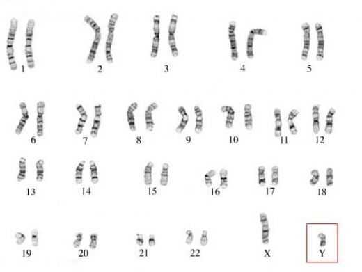
Karyotype analysis is the process of analyzing the number, shape, and size of chromosomes in a cell. By examining these characteristics, medical professionals can identify possible genetic disorders and gain insight into the health of a patient. Karyotypes are typically obtained through a blood sample, which is then analyzed using a microscope and special dyes. The results are laid out in a karyotype worksheet, which is used to interpret the data.
Interpreting Karyotype Worksheets
Karyotype worksheets are used to interpret the results of karyotype analysis. The worksheet is organized into columns and rows, with each column representing a different chromosome. The size, number, and shape of each chromosome are indicated in the rows.
[toc]
In order to interpret a karyotype worksheet, medical professionals must look for abnormalities in the number, size, and shape of the chromosomes. Abnormalities may indicate the presence of a genetic disorder or other health issue.
Conclusion
Karyotype analysis is an important tool for medical professionals to assess the health of a patient. By examining the number, shape, and size of chromosomes, medical professionals can gain insight into possible genetic disorders and the overall health of the patient. In this comprehensive guide, we have explored the basics of karyotype analysis and provided guidance on how to interpret karyotype worksheets.
How to Incorporate Karyotype Worksheet Answers Key Into Your Biology Curriculum
Incorporating karyotype worksheet answers key into a biology curriculum can serve as an effective way to help students gain an understanding of the structure and function of chromosomes. In order to do so, there are several steps that can be taken.
First, the instructor should provide students with an overview of the karyotype worksheet answers key. This overview should include a brief introduction to the concepts of chromosomes, karyotypes, and the use of the key. Additionally, the instructor should explain the purpose of the worksheet and the different types of karyotypes that it can identify.
Next, the instructor should provide students with a worksheet that covers the different types of karyotypes that they may encounter. The worksheet should include a detailed explanation of the different types of karyotypes, including their structure and function. It should also include an explanation of the key for identifying each type.
Finally, the instructor should go over the worksheet with the students, providing them with feedback and guidance as needed. This will help the students become familiar with the concepts and the key, and will also provide them with an opportunity to practice what they have learned.
Incorporating karyotype worksheet answers key into a biology curriculum can be an effective and engaging way to reinforce the concepts of chromosomes and karyotypes. By providing students with a clear understanding of the key and the different types of karyotypes, the instructor can help ensure that students understand the material and are able to apply it to their own studies.
Deciphering the Complexity of Karyotype Worksheet Answers Key: A Step-by-Step Guide to Understanding Chromosome Abnormalities
Step 1: Introduction to Karyotype and Chromosome Abnormalities
Karyotyping is a procedure for determining the number and structure of chromosomes in a cell. It is used to diagnose and evaluate genetic disorders, as well as to detect chromosomal abnormalities. Chromosomal abnormalities can occur due to a variety of reasons, and they may range from small changes in the size or shape of one chromosome to major changes in the number of chromosomes in a cell.
Step 2: Anatomy of a Chromosome
A chromosome is made up of two chromatids, which are joined together at the centromere. Chromosomes are composed of DNA and proteins, and they contain the genetic information that an organism needs to develop and function. Chromosomes are divided into two sections: the short arm (p arm) and the long arm (q arm). The short arm contains a centromere, while the long arm does not.
Step 3: Karyotyping
Karyotyping is the process of examining chromosomes in a cell in order to identify any abnormalities. During this process, stained chromosomes are arranged according to size and shape, and the resulting karyotype is compared to a normal karyotype. Abnormalities can be identified by comparing the chromosomes in the karyotype to a normal karyotype.
Step 4: Types of Chromosomal Abnormalities
Chromosomal abnormalities can be classified into three main categories: numerical abnormalities, structural abnormalities, and mosaicism. Numerical abnormalities refer to an increase or decrease in the number of chromosomes in a cell, while structural abnormalities involve changes in the shape or structure of one or more chromosomes. Mosaicism occurs when the cells in a karyotype contain both normal and abnormal chromosomes.
Step 5: Causes of Chromosomal Abnormalities
Chromosomal abnormalities can be caused by a variety of factors, including genetic mutation, environmental factors, and chromosomal rearrangements. Genetic mutations can be inherited from a parent, or they can arise spontaneously during the development of a cell. Environmental factors, such as radiation or certain chemicals, can also cause chromosomal abnormalities. Chromosomal rearrangements can occur when pieces of chromosomes break off and reattach to other chromosomes.
Step 6: Diagnosis and Treatment of Chromosomal Abnormalities
Chromosomal abnormalities can be diagnosed through a variety of methods, including karyotyping, fluorescent in situ hybridization (FISH), and array comparative genomic hybridization (aCGH). Treatment of chromosomal abnormalities depends on the type and severity of the abnormality. In some cases, doctors may suggest lifestyle changes or medical therapies to manage the symptoms of the disorder. In other cases, surgery or genetic counseling may be recommended.
Step 7: Conclusion
Karyotyping is an essential tool for diagnosing and evaluating genetic disorders, as well as for detecting chromosomal abnormalities. By understanding the anatomy of a chromosome, the process of karyotyping, the types of chromosomal abnormalities, and the causes and treatments available, healthcare professionals can provide the best care possible for patients with chromosomal abnormalities.
Conclusion
The Biology Karyotype Worksheet Answers Key is a useful tool for exploring the fundamentals of karyotyping and chromosome analysis. By studying the answers provided, students can gain a better understanding of the structure and function of chromosomes, as well as the importance of genetic testing. Through this worksheet, students can also gain insight into how abnormalities in chromosome number or structure can lead to genetic disorders. With this knowledge, they can be better equipped to understand the implications of genetic testing and its importance in modern medicine.
[addtoany]
5 photos of the "Biology Karyotype Worksheet Answers Key"
Related posts of "Biology Karyotype Worksheet Answers Key"

Fundamental Counting Principle Worksheet
Exploring the Benefits of Using a Fundamental Counting Principle Worksheet in the ClassroomThe Fundamental Counting Principle worksheet is an invaluable tool for teachers who are looking to enhance their students’ critical thinking skills. This worksheet provides an intuitive and engaging way for students to understand the basics of counting and combinatorics. By engaging in the...

Proving Lines Parallel Worksheet Answers
Exploring the Mathematics Behind Proving Lines Parallel Worksheet AnswersExploring the mathematics behind proving lines parallel is an educational exercise that provides a valuable opportunity to understand the fundamental principles of geometry. In this worksheet, students will learn how to use a variety of tools, such as transversals, angles, and slopes, to prove that two lines...

Surface Area And Volume Worksheet
How to Prepare an Effective Surface Area and Volume Worksheet. 1. Gather necessary materials for the worksheet. This includes a ruler, a calculator, graph paper, and an appropriate textbook. 2. Choose a topic related to surface area and volume. This could include the formulas for calculating surface area and volume, calculating the surface area and...
Circulatory System Worksheet Pdf
Exploring the Benefits of Utilizing a Circulatory System Worksheet PDF in the ClassroomThe utilization of a circulatory system worksheet PDF in the classroom can be a beneficial aid to teaching and learning. A circulatory system worksheet PDF is an interactive educational material, which can be used to help students explore the body’s complex network of...